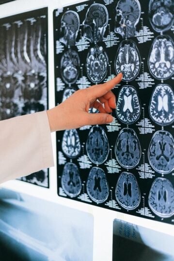
The brainstem is a complex structure that connects the cerebrum of the brain to the cerebellum and spinal cord. It is comprised of three parts: 1) the midbrain, 2) the pons and, 3) the medulla oblongata.
- In the upper posterior portion of the midbrain, one can find the tectum, which has a paired structure named the superior and inferior colliculi. Visual processing and eye movements are within the realm of the superior colliculi, while the inferior colliculi are responsible for auditory processing.
- The pons provide a pathway for tracts to travel up and down our cerebrum and spinal cord. Many nuclei for cranial nerves live in this portion of the brainstem. Motor control involving eye movement and balance/equilibrium maintenance originates here.
- The medulla oblongata holds the nucleus of the solitary tract. This is pivotal for our survival as it reads information about our vital systems, such as our cardiovascular and respiratory systems. It also ensures that we have the reflexive actions to regulate these within a healthy and normal range.
Somatosensory, visual and vestibular sensation information is constantly performing in various areas of our brain to maintain appropriate posture-gait control. Descending pathways from the brainstem to the spinal cord are involved with postural reflexes, head-eye coordination, proper body segment alignment and postural muscle tone. The cognitive process of postural control (which relies on our self-body knowledge, for example, body schema and body motion in space), is significant for our vertical posture. This originates in the temporoparietal association cortex. Seeing as the brainstem and cerebral cortex hold a reciprocating relationship, we need both to be working optimally to maximise the function of our automatic and cognitive processes of posture gait.
We depend on sensory signals coming from both external stimuli and internal visceral information to identify and stabilise negative postural changes. Situated in the centre of the brainstem and expanding from the rostral midbrain to the caudal medulla, lies a complex network of circuits called the reticular formation. The reticular formation consists of a clutter of interdigitating axon bundles with neuron clusters strewn amongst it. These neurons hold a number of functionalities, such as: cardiovascular regulation, control of sensory motor reflexes, sleep management, eye movement organisation and motor control. The descending motor control pathways from the reticular formation impacts the local circuit neurons that manage and organise axial and proximal limb muscles. It is therefore important for our posture and keeping us upright.
The reticular formation delivers information involving the maintenance of our posture in response to any environmental or self-generated disturbances that can have an effect on our body’s position and stability to our spinal cord. Motor centres in the cortex or brainstem control the motor centres in the reticular formation. It ensures that the appropriate neurons in the reticular formation are conducting adjustments that promote the stabilisation of our posture during ongoing movements.
Regarding the significance of visual stimuli in relation to our posture, it is important to incorporate neurological exercises that promote visual training so that we can improve and stabilise dysfunctional neurological outputs that control our postural orientation. We now know that adhering to visual neurological exercises that stimulate our brain stem is extremely beneficial due to the significant role the brain stem plays with maintaining the right posture.
Visual training will help improve control of our visual systems. This will lead to a more efficient orientation of our body within our environment, as well as proper head posture. We can expect to see better results with postural correction when we incorporate an aspect of postural neurology, which will help strengthen these processes, keep us upright and provide some extra love to our brain!

